First-of-its-kind pressurization test could improve Ross procedure outcomes
A new surgical technique includes a pressurization test on a pulmonary valve before it takes on increased blood pressure as an aortic valve.
As a department, we are dedicated to innovation — solving difficult problems others haven't been able to solve and tackling the most complex cases — all while keeping patient comfort and experience at top of mind. Our team continues to use our experience and science to find ways to improve the lives of children and adults with congenital heart disease.
We create new technologies and surgical techniques so that patients can recover as quickly as possible. For example, we invented a heart valve that could reduce the operations a child will need. And we use 3-D imaging to create models of a patient’s heart to prepare a customized surgical plan.
Here are some other innovations that allow us to take on the most complex cases of heart disease and give our patients the best possible outcomes so they can lead healthy lives:
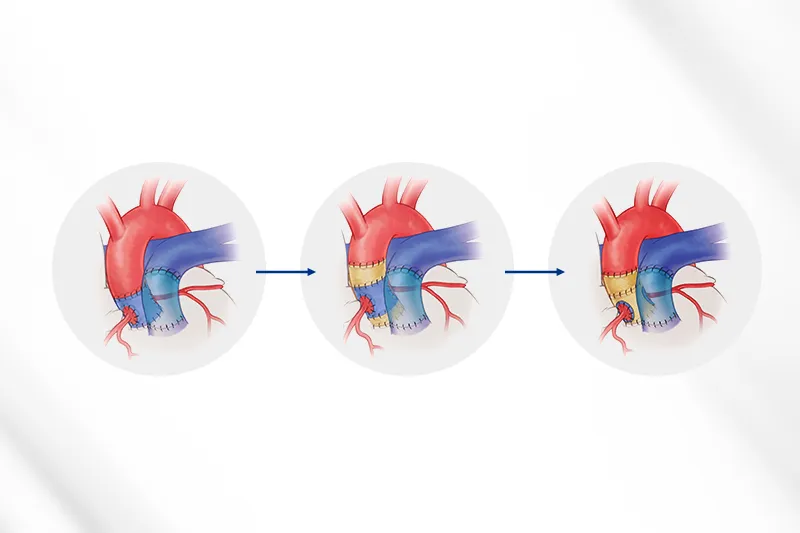
A new surgical technique includes a pressurization test on a pulmonary valve before it takes on increased blood pressure as an aortic valve.
Boston Children’s treats the rare pulmonary disease with a specialized surgery and interventional cardiology procedures.
Ten years’ worth of biventricular repair and Fontan procedure outcomes could help shape future surgery plans.
Ventricular pressure-volume (PV) loop calculations inform decisions about left-ventricle surgeries.

New research by Boston Children’s validates an innovative approach to mapping the heart’s invisible conduction tissue during surgery.

Cardiac surgeons, led by Sitaram Emani, MD, have been testing it as a way to help wean children with congenital heart disease and ischemia-reperfusion injury off ECMO.

Our Heart Center team performed this procedure on a 4-year-old boy, implanting a donor’s aortic valve.

The Boston Children’s cardiac surgery team is seeing lasting positive outcomes in patients after making adjustments over the years to the Ross procedure, a last-option treatment for aortic valve disease.

Aiming to reduce the need for pacemakers, Boston Children’s clinicians have created two new approaches to cardiac surgery that can identify elusive conduction tissue and potentially decrease heart block.

Three-dimensional modeling allows our team of experts to capture a comprehensive view of a child’s heart before surgery and plan the most effective repair procedure.

Boston Children’s is the first hospital in the world to use this procedure as part of a strategy to surgically rehabilitate the left ventricle in patients with borderline left heart.

Our heart specialists leaned into technological innovation, their experience, and a perseverance that would ultimately confirm their belief that even the highest-risk cases are not out of reach.

Our cardiac surgeons found that using confocal microscopy could reduce arrhythmia in congenital heart defect operations.

Boston Children's surgeons' new technique allows for the consistent identification of conduction tissue in complex cases of CHD.

Surgeons at Boston Children's were inspired by Chinese finger traps.

Training heart surgeons in the procedure for children provides an option for countries that cannot access mechanical aortic valves.

An airway stent, custom-fit in real time, could benefit patients with tracheobronchomalacia. The design may be adaptable for heart implants.